Monday, April 17, 2017

Cystic Fibrosis


CYSTIC FIBROSIS 

Related image

The day of convention at Washington Adventist University. There were several presentations  that caught my attention, because most of them involve some type of disease. There was one specific that I appreciated. My friend and classmate made an impressive model of a pair of lungs, one had purple flowers that represent the awareness for cystic fibrosis and the other one affected lung of the disease. The information that she presented was an oral presentation and she went over what cystic fibrosis was all about. Cystic fibrosis is a genetic disorder that affects the lungs but can also affect other organs such as the pancreas, liver, and kidneys. Some of the symptoms that are presented are sinus infections, difficulty breathing, and clubbing of fingers and toes. Cystic fibrosis is an inherited autosomal recessive disease, that causes by the presence of mutations. She also mentioned that unfortunately there is no cure for this condition, but it can be treated with antibiotics.In some cases lung transplantation is necessary in the case the lungs continues to deteriorate. I thought she did a very good job presenting the cell and molecular aspect of this disease.

Parkinson Disease

Investigation


Parkinson's Disease 

Parkinson's sickness is a constant and dynamic development issue, implying that side effects proceed and decline after some time. Almost one million individuals in the US are living with Parkinson's sickness. The cause is obscure, and in spite of the fact that there is no cure, there are treatment choices, for example, prescription and surgery to deal with its side effects.

Parkinson's includes the glitch and passing of imperative nerve cells in the brain, called neurons. Parkinson's principally influences neurons in a region of the cerebrum called the substantial nigra. Some of these withering neurons deliver dopamine, a substance that sends messages to the piece of the cerebrum that controls development and coordination. As Parkinson’s  advances, the measure of dopamine delivered in the cerebrum diminishes, leaving the effects of uncontrollable muscle movement.

Cell Molecular

Parkinson's Disease is "idiopathic"meaning that the caused of it is unknown. There is about a 15 percentage that can cause the disease. A mutation on the gene can cause Parkinson's Disease. A gene that can be affected is chromosome 6 has a mutation in Park 2 gene that can be inherited by an Autosomal Recessive. The park 2 gene is one of the largest gene and it encodes for protein called Parkin. The exact function of parkin is to tag other damaged or excess protein to ubiquitin and be transferred to proteasomes to be breakdown.


Sunday, April 9, 2017

Lysosomes


Image result for lysosome
Lysosomes


Lysosomes are membrane-bound organelles that cut or digest molecules found in a cell. The function of lysosomes can be breaking down into two processes autophagy and crinophagy. Autophagy means self-eating, and it's when lysosomes digest molecules that are part of the cell itself, or other cells. If there are organelles that are no longer functional, these will be digested by lysosomes. A good example of autophagy would be the macrophages of the immune system. Macrophages function its to engulf bacteria and viruses and then be transferred to the lysosomes to digest. Crinophagy is when the lysosomes digest excess secretory products. An example of crinopahgy will be if a cell is producing too much of a hormone, lysosome will break down the extra hormone that it's being produced. In both autophagy and crinophagy, after the lysosomes complete the breakdown of molecules. It will release the molecules into the cytoplasm for reused. 

Saturday, April 1, 2017

Viruses vs Cancer


Cancer vs Virus 

Cancer is a group of diseases that characterize by abnormal and uncontrolled cell proliferation. Cancer cells growth with the potential to invade and interfere with other organs systems functioning. The initiated cause for cancer it comes from a mutation in the genes that usually control cell proliferation. The identification of the mutating genes came from the studies of viruses that cause cancer in animals. Furthermore, studies eventually were able to identify the specifics of cancer or the causing gene carried by the virus. And what is the relationship with normal cells? Although some cancers are known be caused by a virus; some occur from mutations in normal cells genes. There are studies of cancer-causing viruses, which had led to the identification of a various gene responsible for nonvirus induced cancer, this has helped to understand the molecular mechanisms responsible for cancer. In the search for a cancer treatment, these insights into the molecular and cellular biology are important to achieve this step.

Source:
"Viruses That Can Lead to Cancer." American Cancer Society. The American Cancer Society Medical and Editorial Content Team, 11 July 2016. Web. 011 Apr. 2017.


Sunday, March 26, 2017

Cytoskeleton Microtubules


Cytoskeleton Microtubules 
Related image

The cytoskeleton is composed of protein fibers that determine the shape of a cell without cell walls. It primarily provides for movement of cells, for the structural support, movement organelles within the cell and the framework for moving and separating chromosomes during cell division. The three types of protein fibers in the cell are thick microtubules,  thin microfilaments, and medium sized intermediate filaments. Microtubules structure are hollow tubes that assemble from subunits made out of the protein tubulin. Cell subunits are assembled into microtubules by microtubule organizing centers found in many locations inside the cell, in particular, eukaryotic cells have a microtubule organizing center near the nucleus which is known by the name of the centrosome.  The centrosome during cell division span the particular microtubes call the spindle apparatus which separates chromosomes and divides then between the two new daughter cells.

Sunday, March 19, 2017

Natural Death


Reflection 


Natural Death 
Image result for apoptosis


I came across an interesting article titled: "What Our Cells Teach Us About a Natural Death." The author Haider Javed Warraich compares and contrast two different points the deterioration of cells and the end of life. At the begging of his article, he sets scenarios and explains how death has been view throughout history. He then makes a transition to the end cycle of cells. The author explains that cell dies via three primary mechanisms. One which is considered the ugliest form of cell death is necrosis; it occurs because of lack nutrients, cell's burst open by this releasing their content. The second mechanism of cell death is called autophagy, and this is when a cell turns on itself which the redundant cytoplasmic components transform into lysosomes. The third and most known cell death is apoptosis, is the natural deterioration or signaling molecules undergo a form of self-demolition. As a result, apoptosis is convenient for ourselves; it regulates the structures and growth of our organisms. At the end of the article, the author makes his point of viewed. What is beneficial for an organism is best for the cell, which whether a cell has to die in appropriate fashion for the sake of society, ecosystem, or body.

Source: 
Warraich, Haider Javed. "What Our Cells Teach Us About a ‘Natural’ Death." The New York Times. The New York Times, 13 Mar. 2017. Web. 19 Mar. 2017.

Friday, March 3, 2017

Kidney Stones

Encounter

Kidney Stones Formation

I decided to write about Kidney Stones because a family member has encounter this condition. I got a called this week from my father and in our conversation he mentioned his excruciating pain in his lower back. As Biology major I started to put his symptoms to figure out what was the cause from his pain. I didn't imitatively know what it was, but I did my researcher on the possibilities of his pain and everything pointed out to kidney stones. I told my dad to not take my word for it and I then encourage him to see a physician to get tested. Half way through the week my father calls me and tells me that I was right, it was kidney stones. The doctor than prescribe him medicine for the discomfort and expulsion of the stones. I wanted to be right, I didn't wanted to be something worst. I was than intrigue and curious on how this small components are form. Here is what I found out. 

I kidney stones are easily formed, some even can be the size of a head pin or a small rocks. The pain resulting from this crystal structure can be dangerous. Another name for kidney stones is renal calculi, are aggregates of crystals enclosed in a matrix that is developed inside the kidney. There are five major categories of crystals which are: calcium oxalate, calcium phosphate, struvite, uric acid, and cysteine. The most common crystal is calcium oxalate. For a stone to form it has to go through several phase. The first phase is nucleation, ions like calcium and oxalate are filter into the urine by the kidney spontaneously and then join together to form a solid crystal. The crystal then travels along nephron and then deposited at the renal papilla where they obtain grow and this is the second phase. Crystals that are in the same place and have already form stick together with other crystals to create a clusters this is the third phase aggregation. The last phase is retention, The clusters than are formed to a stone that which are retain in the kidney where they continue growing for an unspecified time. Then the stones are moved and are displaced into the ureter tube. If the stones continues to grow until it reaches a critical size it can be too large to pass easily through  the ureteropelvic junction, the illiac artery, or the ureterovesicle junction. In result there is pain and obstruction, until the stone slowly passes into the bladder and then expelled through the urine stream.  

Source :Evan, Andrew P. "Physiopathology and Etiology of Stone Formation in the Kidney and the Urinary Tract." Pediatric Nephrology (Berlin, Germany). Springer-Verlag, 01 May 2010. Web. 03 Mar. 2017.

Friday, February 24, 2017

Exoplanet Kepler-186 F


Reflection

Exoplanets Discovery  

Kepler-186 F 

Related image

       The exoplanet Kepler-186 F was named after NASA's Kepler Space Telescope. Kepler is the first earth size planet discovered in the habitable zone orbiting a star. Astronomers believed that Kepler is at the correct distances from a star that can indicate liquid water might pond on the surface of the planet. Many other exoplanets have being found in the habitable zone before, but most of this planets were at least 40 percent larger than Earth. Kepler-185 F in the other hand is more reminiscent of Earth. Although Kepler size is known, there is still a lot more to be discovered like it's mass and composition. The exoplanets discovered are approximately 500 light-years from Earth and orbit around a star which is half the size and mass of our sun. Even though it will be far long for human kind to find or travel to another habitable planets, astronomers theorized the possibilities that maybe future generations will. For right now searching for life outside the solar system is not a priority, but finding planets with the same characteristics that mimic that Earth is the main priority after this extraordinary discovery.  

Source: Culler, Jessica. "Kepler-186f, the First Earth-size Planet in the Habitable Zone." NASA. NASA, 03 Mar. 2015. Web. 24 Feb. 2017.

Sunday, February 19, 2017

Enzymes of Recombinant DNA

Terminology 

A. Nucleases - Enzyme that cleaves the chains of nucleotides into smaller units.
     1. Exonuclease- An enzyme that digests nucleic acids starting from the 5' or 3' terminus.
     2. Endonuclease- An enzyme that digests nucleic acids from within the sequence.
     3. Isoschizomer- Restriction endonucleases that contain an identical site but it can be derived from different species of bacteria.
B. Polymerases- Enzyme that brings about the formation of a particular polymer, especially DNA or RNA.
     1. DNA Polymerases- An enzyme that synthesizes DNA from a DNA template.
     2. RNA Polymerase II - The enzyme that is used to transcribe structural genes that result in mRNA.
    3.  RNA Polymerase III- The enzyme that is used by the cell to transcribe ribosomal RNA genes.
    4. Kinases- Enzymes that transfer the Y-phosphate group from ATP to the 5' hydroxyl group of a nucleic acid chain.
    5. Ligases- The enzyme that utilizes the Y-phosphate group of ATP for energy to form a phosphodiester linkage between two pieces of DNA.
    6. Reverse transcriptase- Enzyme that purifies first from retrovirus-infected cells, to produce a cDNA copy from an mRNA molecule.
    7. Telomerase - A specialized DNA Polymerase that protects the length of the terminal segment of a chromosome.


Monday, February 13, 2017

Nuclear Transport

Investigation

Cell Nuclear Exportin

                           Image result for nuclear exportin
The nuclear import complex it controls the entering and exit of large molecules through the nucleus pores. Small proteins pass easily through the nuclear pore, as for large proteins, it is more complex to enter. The transport of large proteins in either direction requires various steps. Exportin is a protein that is found in the nucleus, which it helps with the transportation of materials from the nucleus to the cytosol. For exportin to function properly, it needs the help other molecules. Exportin bounds to Ran-GTP, which eventually will cause a conformational change in exportin that causes the attachment to the nuclear export signal. The nuclear export signal is a short amino acid sequence also known as the cargo protein. Once all of these three components are attached together, these will travel than travel through the nuclear pore complex. In the cytosol, GTPase will start hydrolyzing Ran-GTP into Ran-GDP which cause the releasing of Ran-GDP fro exportin. Exportin can repeat the process again, by detaching from the cargo protein. Next exportin it travels back to the nucleus and the cargo protein stays in the cytosol. 

Resource 
"Nuclear Transport." Nuclear Transport. N.p., n.d. Web. 11 Feb. 2017.

Sunday, February 5, 2017

Human or Pig?

Research 

 Human?Or Pig?
Scientists Create Human-Pig Hybrid In The LAb
The power of science and medicine has come to another discovery. Scientists have presented the first successful human-animal hybrid. The project proves that the human cells can be introduced into a non-human organism to survive, and even grow inside a host animal, in this case, pigs. In the past, the United States have rejected to provide funds to projects like this one. The public opinion as well has been against the creation of organisms that are half human and half animal. The image above is a pig embryo that was injected with human cells. scientists are currently monitoring the developments of a four-week-old embryo. So far there is a lot of controversies if this project should continue or should be stopped. In the other hand, if this project is a success this could be a game changer for organ transplants in the coming future. 

Source: Blakemore, Erin. "Human-Pig Hybrid Created in the Lab-Here Are the Facts." National Geographic. National Geographic Society, 27 Jan. 2017. Web. 04 Feb. 2017.

Friday, January 27, 2017

Benefits of Green Tea

Reflection  

Columbia Journal 
Biology and Chemistry section 

The New Western Craze 
by: Tayde Quinto 

Image result for green tea

Green Tea has proven to be the most popular drink in the world. In the article, the writer mentioned that people that consumed regularly green tea have a lower risk of having stress, anxiety, and depression. An example of green tea referred to in the article was Camellia sinensis, this type of green tea is less fermented, and as a result, it contains more antioxidants. A great benefit of green tea are the various effects of prevention of tooth decay, inhibition of high blood pressure, cancer prevention, and others. Another component that green tea contains is caffeine, which can cause increased of alertness, increased stamina, and hangover prevention. In addition to her article, she mentions green tea can also help stabilize blood pressure and cholesterol levels. She finalized by recommending drinking a few cups of green tea every day to experience the health benefits that green tea can offer.    

Friday, January 20, 2017

Cell and Molecular Class

Reflection

Cell and Molecular Class

The molecular cell class it focused on the study of cellular process or the basic unit of life. The information provided its link with biochemistry, immunology, genetics, and microbiology. In detail cell biology is the study of different structures and functions of the cell. Throughout the semester, the class will emphasize on the following: Organization of organelles, life cycle, cell interactions with the environment, metabolic processes, physiological properties, signaling pathways. The first two weeks in the class we have covered microscopic and differentiation between prokaryotic and eukaryotic cells. As a class, we are hoping to learn how cells work and this essential to understand diseases and disorders in the human body, to successfully apply them in our professional careers.
  

Friday, January 13, 2017

Cell and Molecular Terminology

Dictionary 


Microscopes Techniques

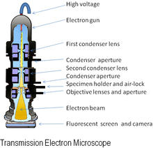
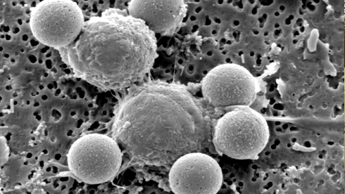
  • EM microscopy- Is the use of an electron microscope that are used study ultra structure inorganic specimens such as: microorganism, cells, biopsy, metals, and large molecules. The microscope uses a beam of electrons as light. The wavelength that it produced it's about 100,000 times smallest than a light microscopes. 
Image result for EM microscopy images      Image result for EM microscopy images
  • Light microscope- Also known as the optical microscope, this type of microscope uses visible light and lenses to visualize images of the samples. The optical microscope has being the initial invention to bring out some other microscope that as well uses light. For example: fluorescence microscopy, dark and bright field, digital microscope. 
Image result for Light microscope and images
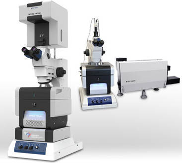
  • Force Spectrum microscopy- It's the application technique of Brownian motion, which is the motion of particles that are suspended in fluid. This can amplify the magnitude of random forces that can interfered with the oscillation frequency of particles when is viewed from a spectrum microscope.
Image result for Force Spectrum microscopy images
  • Polarizing microscope- Another name for the polarizing microscope is petrographic microscope and mainly used for geological specimens, although this microscope has being a great used in medicine and biology. The main component that differentiates from other microscopes is that uses polarized light, which are light waves traveling in one direction.   
  Image result for polarizing microscope images